
An Ebola outbreak represents a severe public health crisis caused by the Ebola virus, a pathogen that triggers hemorrhagic fever in humans and non-human primates. These outbreaks originate through zoonotic spillover—the transmission of a virus from infected animals to humans—and propagate through direct contact with bodily fluids. For policymakers, health professionals, and agencies based in Washington, understanding the mechanisms of transmission, historical contexts, clinical progressions, and international containment protocols is essential for protecting global health security and domestic biodefense.
- What Is an Ebola Outbreak and How Does the Virus Spread?
- What Is the History of Documented Ebola Outbreaks?
- What Are the Pathological Mechanisms of the Ebola Virus?
- What Are the Clinical Stages and Symptoms of Ebola Virus Disease?
- How Do Public Health Authorities Diagnose and Treat Ebola?
- What Interventions Contained Previous Epidemics?
- What Are the Global Security and Future Policy Implications of Outbreaks?
What Is an Ebola Outbreak and How Does the Virus Spread?
An Ebola outbreak is a sudden increase in cases of Ebola virus disease within a human population, initiated by animal-to-human transmission and sustained through direct contact with the blood, secretions, organs, or other bodily fluids of infected individuals.
The Ebola virus belongs to the family Filoviridae and genus Ebolavirus. The genus consists of six distinct species, including Zaire ebolavirus, Sudan ebolavirus, Tai Forest ebolavirus, Bundibugyo ebolavirus, Reston ebolavirus, and Bombali ebolavirus. Only the first four species cause severe disease in humans. The natural reservoir host remains the fruit bat, specifically species from the chapters Hypsignathus, Epomops, and Myonycteris.
Spillover occurs when humans handle or consume infected bushmeat, such as fruit bats, chimpanzees, gorillas, monkeys, forest antelopes, and porcupines. Once the virus enters a human host through broken skin or mucous membranes, human-to-human transmission sustains the outbreak.
Transmission requires direct contact with infected bodily fluids. These fluids include blood, saliva, vomit, faeces, sweat, semen, breast milk, and urine. The virus is not airborne. It does not spread through casual contact, water systems, or routine respiratory droplets.
The environment plays a significant role in transmission dynamics. The virus remains infectious on solid surfaces, including medical equipment, bed linens, and clothing soiled with bodily fluids, for several days at room temperature. Traditional burial practices involving direct contact with the deceased body contribute significantly to amplification during outbreaks.
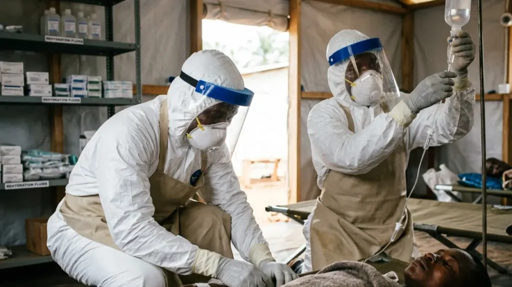
Healthcare settings frequently experience nosocomial transmission—infections acquired within a hospital—when staff members work without adequate personal protective equipment (PPE) or fail to implement strict infection prevention and control (IPC) measures.
What Is the History of Documented Ebola Outbreaks?
The history of documented Ebola outbreaks began in 1972 with a retrospective case and gained formal recognition in 1976 through simultaneous outbreaks in the Democratic Republic of the Congo and Sudan, followed by the catastrophic 2014 West African epidemic.
The first recognized outbreaks occurred in 1976. The first outbreak emerged in Yambuku, a village near the Ebola River in the Democratic Republic of the Congo (then Zaire). This outbreak involved the Zaire ebolavirus species, resulting in 318 cases and 280 deaths, yielding a case fatality rate of 88 percent.
The second 1976 outbreak occurred concurrently in Nzara, Sudan (now South Sudan). Caused by the Sudan ebolavirus species, this event led to 284 cases and 151 deaths, establishing a case fatality rate of 53 percent. Subsequent decades saw sporadic, localized outbreaks across Central Africa. Significant events occurred in Kikwit, Democratic Republic of the Congo, in 1995 (315 cases, 254 deaths) and in Gulu, Uganda, in 2000 (425 cases, 224 deaths).
The largest outbreak in history was the 2014–2016 West African epidemic. It began in a rural village in Guinea in December 2013 and rapidly crossed land borders into Liberia and Sierra Leone. This epidemic transformed the disease from a localized rural crisis into an urban phenomenon, affecting major metropolitan areas including Conakry, Monrovia, and Freetown.
By the time the World Health Organization (WHO) declared the end of the epidemic in June 2016, the virus had infected 28,616 people and caused 11,310 confirmed deaths across eleven countries, including localized transmissions in the United States, Spain, and Mali. This widespread disruption prompted emergency response operations from federal agencies headquartered in Washington to manage domestic monitoring and international aid.
Subsequent major outbreaks returned to Central Africa. The 2018–2020 Kivu outbreak in the eastern Democratic Republic of the Congo became the second-largest occurrence on record. Operating within an active conflict zone, healthcare workers faced armed resistance, resulting in 3,470 cases and 2,280 deaths. In 2022, Uganda experienced an outbreak of Sudan ebolavirus, comprising 164 cases and 55 deaths, illustrating the persistent risk of different viral strains.
What Are the Pathological Mechanisms of the Ebola Virus?
The pathological mechanisms of the Ebola virus involve the systematic destruction of the host immune system, widespread endothelial cell damage causing vascular leakage, and severe disseminated intravascular coagulation that leads to multi-organ failure and profound hypovolaemic shock.
Pathogenesis begins when the viral glycoprotein attaches to host cell receptors, specifically targeting immune cells such as macrophages, monocytes, and dendritic cells. Upon entry, the virus replicates rapidly, suppressing the host’s initial immune response. The viral protein VP24 blocks interferon signalling, preventing the host cell from mounting an antiviral defense, while viral protein VP35 inhibits the activation of regulatory proteins that signal immune responses.
As infection progresses, the virus destroys these target immune cells, triggering an uncontrolled release of pro-inflammatory signaling proteins called cytokines. This phenomenon, known as a cytokine storm, includes elevated levels of interleukin-1, interleukin-6, and tumour necrosis factor-alpha. The cytokine storm increases the permeability of endothelial cells—the cells lining the interior surface of blood vessels.
Simultaneously, the virus migrates to the liver, spleen, and lymph nodes via the lymphatic system. In the liver, the virus infects hepatocytes, destroying the organ’s ability to produce clotting factors. This structural breakdown initiates disseminated intravascular coagulation (DIC), a condition where small blood clots form throughout the bloodstream, blocking small blood vessels and depleting platelets needed for normal clotting.
The combination of endothelial damage, vascular leakage, and coagulation failure produces the classic symptoms of Ebola hemorrhagic fever. Blood leaks from the vascular system into surrounding tissues and body cavities. Severe fluid loss occurs primarily through gastrointestinal tract manifestations, including continuous vomiting and profuse diarrhea. This massive loss of fluids induces hypovolaemic shock—a fatal drop in blood pressure and circulating blood volume—culminating in multi-organ failure, particularly affecting the kidneys, liver, and lungs.
What Are the Clinical Stages and Symptoms of Ebola Virus Disease?
The clinical stages of Ebola virus disease progress rapidly from an initial asymptomatic incubation period to acute flu-like symptoms, followed by severe gastrointestinal dysfunction, systemic vascular leakage, multi-organ breakdown, and either death or prolonged convalescence.
The incubation period—the time elapsed between exposure to the virus and the first appearance of symptoms—ranges from 2 to 21 days, with an average duration of 8 to 10 days. Individuals are not contagious during the incubation period.
Stage 1: The Dry Phase
The illness begins abruptly with the “dry phase,” characterized by non-specific systemic symptoms. Patients experience a sudden onset of high fever, exceeding 38.3°C (101°F), accompanied by profound asthenia (severe physical weakness), severe headache, generalised myalgia (muscle pain), arthralgia (joint pain), and a painful sore throat. This stage lasts approximately one to three days and mimics other tropical endemic diseases, including malaria, typhoid fever, and dengue fever.
Stage 2: The Wet Phase
Between days three and seven, the disease transitions into the “wet phase,” defined by severe gastrointestinal distress. Patients suffer from intractable nausea, projectile vomiting, and voluminous, watery or bloody diarrhea. The fluid loss during this stage is extreme, often exceeding five to ten litres of fluid per day. Abdominal pain is sharp and continuous.
Stage 3: Advanced Systemic Illness
By day seven to ten, the disease reaches its advanced systemic stage. Metabolic acidosis and severe electrolyte imbalances, such as hypokalemia (critically low potassium) and hyponatremia (critically low sodium), disrupt cardiac and neurological functions.
Maculopapular rash—a flat, red area on the skin covered with small confluent bumps—often develops on the trunk and extremities. Hemorrhagic manifestations occur in approximately 40 to 50 percent of cases. These include:
- Internal hemorrhaging: Bleeding within the gastrointestinal tract, lungs, and pleural cavities.
- External hemorrhaging: Petechiae (small red spots on the skin caused by minor bleeding), ecchymosis (bruising), bleeding from venipuncture sites, epistaxis (nosebleeds), and hematemesis (vomiting blood).
Neurological involvement manifests as confusion, delirium, convulsions, and coma. Death typically occurs between days six and sixteen from hypovolaemic shock and multi-organ failure. Patients who survive show clinical improvement by day ten, though they encounter a prolonged convalescence period marked by uveitis (eye inflammation), orchitis (testicular inflammation), severe arthralgia, and significant fatigue.
How Do Public Health Authorities Diagnose and Treat Ebola?
Public health authorities diagnose Ebola using molecular testing methods, specifically reverse transcription-polymerase chain reaction assays, and treat the disease through specific monoclonal antibody therapies combined with aggressive, protocol-driven intravenous fluid and electrolyte resuscitation.
Early diagnostic confirmation is critical for containment. Because early symptoms mirror other febrile illnesses, laboratory diagnosis is required. The gold standard diagnostic tool is the reverse transcription-polymerase chain reaction (RT-PCR) assay. This molecular test detects viral ribonucleic acid (RNA) in blood specimens.
Viral loads are typically high enough for detection within three days of symptom onset. For safety, laboratories also utilize enzyme-linked immunosorbent assays (ELISA) to detect specific viral antigens or IgM and IgG antibodies, alongside rapid diagnostic tests (RDTs) for screening in remote field environments.
Therapeutic interventions advanced substantially following clinical trials conducted during the 2018–2020 Kivu outbreak. The U.S. Food and Drug Administration (FDA), an executive agency based in Washington, approved two specific monoclonal antibody treatments for Zaire ebolavirus:
- Inmazeb (atoltivimab/maftivimab/odesivimab-ebgn): A combination of three monoclonal antibodies that bind to different epitopes on the viral glycoprotein, neutralizing the virus.
- Ebanga (ansuvimab-zykl): A single monoclonal antibody that prevents the virus from binding to the host cell receptor.
These therapeutics significantly reduce mortality rates when administered early in the course of the disease.
Concurrently, supportive care remains the cornerstone of survival. Aggressive intravenous fluid administration is mandatory to counter gastrointestinal losses. Clinicians infuse balanced crystalloid solutions, such as Ringer’s lactate, guided by regular measurement of serum electrolytes, blood glucose, and acid-base status.
Symptomatic management includes antiemetics to control vomiting, analgesics for pain management, and broad-spectrum antibiotics if secondary bacterial infections or translocation of gut bacteria occur due to intestinal wall damage.
What Interventions Contained Previous Epidemics?
The interventions that successfully contained previous epidemics comprise rapid isolation of infected individuals, rigorous contact tracing, community-engaged safe and dignified burials, ring vaccination strategies, and the strict enforcement of infection prevention protocols within healthcare facilities.
Epidemic control relies on breaking the chain of transmission through a multi-pillar public health framework. The primary intervention is rapid isolation and treatment. Dedicated Ebola Treatment Centres (ETCs) isolate confirmed patients from the general population, reducing community exposure. These units operate under strict bio-containment rules, utilizing unidirectional traffic workflows and designated zones divided into high-risk and low-risk areas to protect medical personnel.
Contact tracing is the systematic identification and monitoring of persons exposed to a confirmed case. Contact tracers identify three rings of contacts: primary contacts (direct exposure), secondary contacts (exposed to primary contacts), and tertiary contacts. Trackers monitor these individuals daily for 21 days from their last known exposure, taking their temperature and assessing for early symptoms. If a contact develops a fever, they are moved to an isolation facility immediately.
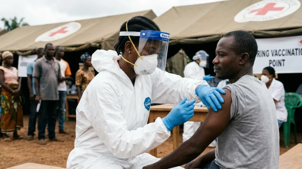
Vaccination strategies changed outbreak dynamics during the late 2010s. The Ervebo vaccine (rVSV-ZEBOV) is a live-attenuated, recombinant vesicular stomatitis virus vectored vaccine expressing the Zaire ebolavirus glycoprotein. Public health agencies deploy this vaccine using a “ring vaccination” strategy:
- First Ring: Vaccinating the immediate primary contacts of a newly diagnosed patient.
- Second Ring: Vaccinating the contacts of those primary contacts (the secondary contacts).
- Healthcare Workers: Vaccinating frontline medical staff and laboratory workers in the affected zone.
This ring vaccination method establishes a human buffer zone of immunity around the virus, preventing further geographic spread.
What Are the Global Security and Future Policy Implications of Outbreaks?
The global security and future policy implications of Ebola outbreaks include the structural collapse of regional economies, the generation of long-term biological security risks, and the clear need for sustained international financing of decentralized surveillance infrastructure.
Ebola outbreaks cause profound damage extending beyond direct mortality figures. Economically, epidemics destabilize entire regions. The World Bank reported that the 2014 West African epidemic caused over 2.8 billion US dollars in lost gross domestic product (GDP) across Guinea, Liberia, and Sierra Leone. Cross-border trade halts, commercial airline flights cancel, and agricultural production drops due to movement restrictions, triggering severe food insecurity.
Healthcare systems collapse under the strain of an outbreak. Resources shift entirely to Ebola response operations, causing routine healthcare services to cease. During the West African epidemic, maternal mortality increased by 111 percent, and infant mortality escalated due to the suspension of routine immunization programs for measles, polio, and tuberculosis. The loss of healthcare workers to the disease exacerbates these system failures permanently.
From a global security perspective, the potential for cross-border introduction via international air travel requires strict implementation of the International Health Regulations (IHR 2005). National governments maintain core capacities for exit and entry screening at international airports, seaports, and land crossings. In Washington, federal legislative bodies and defense agencies continuously evaluate biodefense funding allocations to ensure early warning mechanisms and domestic containment units are fully operational.
Biosecurity policies must regulate the handling of filoviruses in high-containment laboratories (Biosafety Level 4), preventing accidental release or weaponization.
Future policy directives emphasize proactive preparation rather than reactive assistance. The World Health Organization maintains emergency stockpiles of therapeutics and vaccines, but decentralizing this supply chain to regional hubs within Africa remains an ongoing priority for international donors. Policy experts emphasize investing in community-led surveillance systems and basic water, sanitation, and hygiene (WASH) infrastructure in remote areas. This focus ensures early detection and immediate containment before localized spillovers transform into catastrophic international emergencies.